


In brief: A 3D cone beam CT (CBCT) scan gives a three-dimensional view of the teeth, jaws, nerves and sinuses, so dentists can measure bone height, width and density that flat X-rays cannot show. It is the standard planning scan before implants and is also used for complex root canals, wisdom teeth, sinus and jaw joint problems. The dose is low, and a scan is taken only when its answer can change the treatment.
What a CBCT scan is and how it works
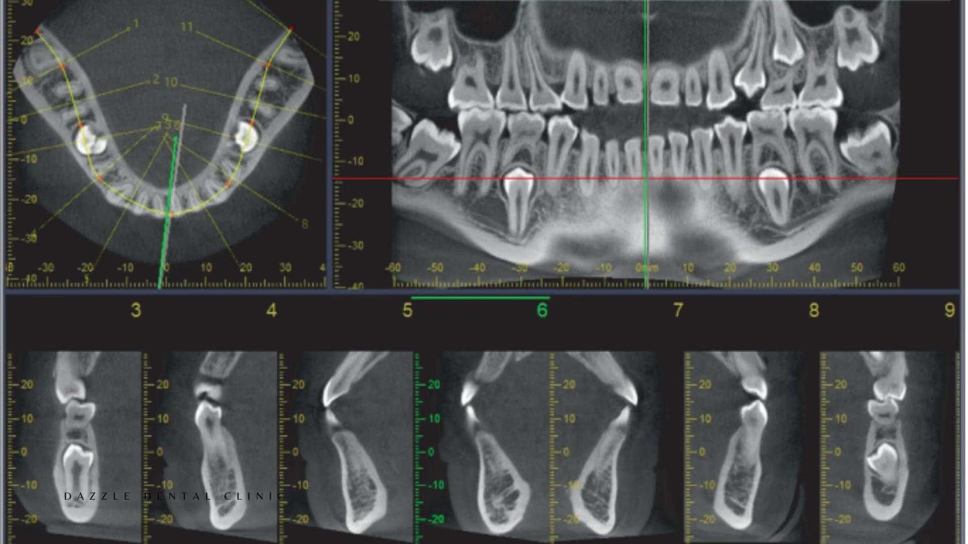
CBCT stands for cone beam computed tomography. A cone-shaped X-ray beam and a detector rotate once around your head, usually in about 10 to 40 seconds, while you sit or stand still. Software rebuilds the hundreds of images into a 3D volume.
The dentist can then slice that volume in any direction: horizontal (axial), front-to-back (coronal) and side-to-side (sagittal) sections, a panoramic view similar to an OPG, and 3D surface images. The whole visit, including positioning, usually takes a few minutes, and the images are ready straight away.
The field of view can be small (one area of the jaw) or large (both jaws and sinuses). The smallest field that answers the clinical question is chosen, because a smaller field means a lower dose.
CBCT vs regular dental X-rays
Periapical X-rays, bitewings and the panoramic OPG are two-dimensional. They show bone height, cavities, gross bone levels and large lesions well, and they remain the right choice for routine check-ups.
Their limit is that depth is lost. Structures in front of and behind each other overlap, so a 2D image cannot show:
- Bone width: a ridge that looks tall enough on an OPG may be too narrow for the implant.
- The outer (buccal) bone plate: the first bone to shrink after extraction or gum disease.
- Exact nerve and sinus position: the lower jaw nerve shows as a line, but not where it sits from cheek side to tongue side.
- Hidden problems: small lesions between roots, root tips left from old extractions, cysts and extra canals.
CBCT for dental implant planning
For implants, CBCT is the core planning tool. At each planned site it measures bone height above the lower jaw nerve or below the sinus floor, the bone width for the implant diameter, and grey-scale density that broadly matches the Misch bone types (D1 to D4). It also locates the mental foramen, nasal floor and any leftover pathology.
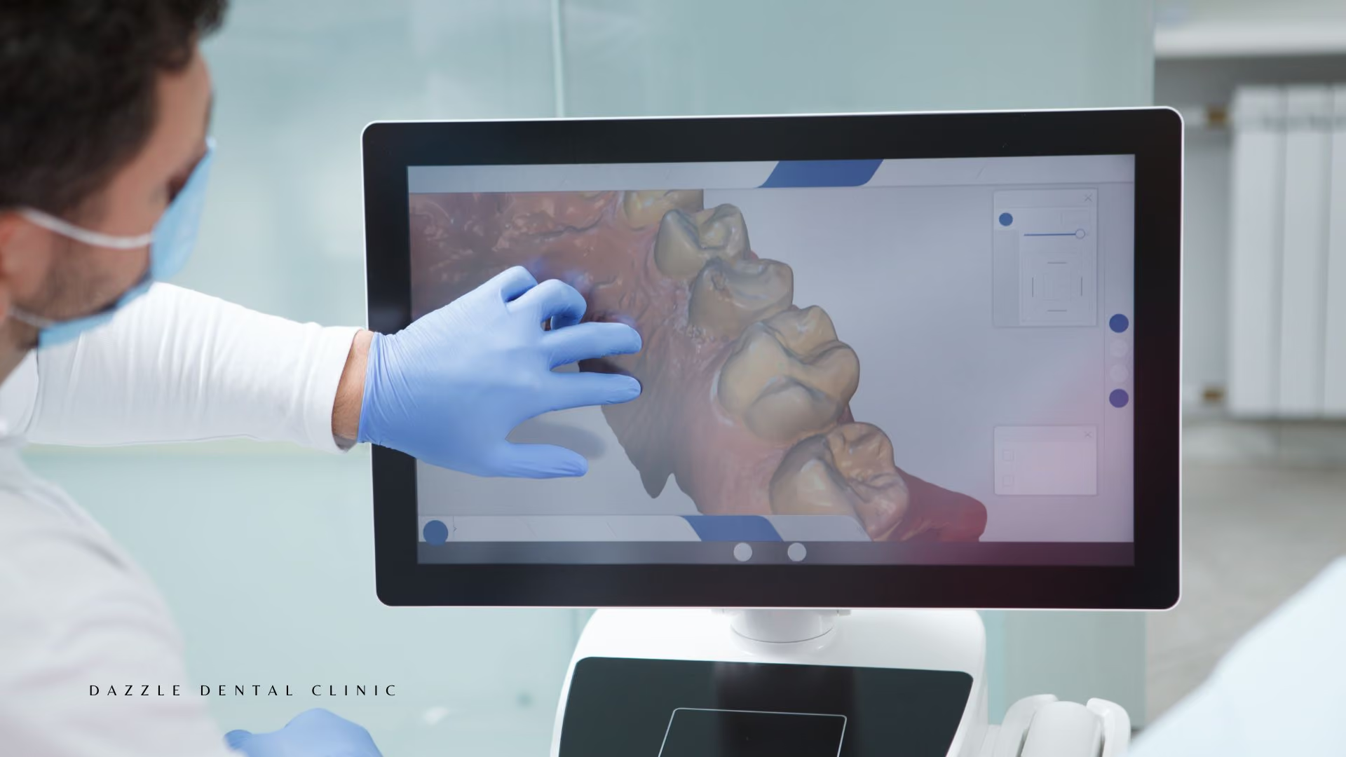
The scan is imported into planning software, often together with a TRIOS 5 intraoral scan of the teeth and bite. A virtual implant is placed where both the bone and the future crown need it. If the position comes too close to a nerve or would break through the bone wall, it is changed on screen before surgery.
That plan then drives surgery: either a 3D-printed surgical guide or, at Dazzle, ClaroNav Navident guided surgery, which tracks the drill against the CBCT in real time. Our article on bone quality and volume assessment explains what the measurements mean.
When a CBCT is essential before implants
- All full-arch cases, such as All-on-4 and All-on-6.
- Upper back teeth, where the sinus is close.
- Lower back teeth near the nerve.
- Moderate or severe bone loss, or a planned graft or sinus lift.
- Immediate implants and same-day teeth.
- Zygomatic and pterygoid implants, where the path runs near the eye socket floor and the sinus. See our guide to zygomatic and pterygoid implants.
Other uses of CBCT in dentistry
- Root canal treatment: shows extra canals (such as the second mesiobuccal canal, MB2, found in more than half of upper first molars in many studies), C-shaped canals, root curves, cracks and the true size of infections at the root tip. It helps when a previous root canal has failed for an unclear reason.
- Wisdom teeth: shows whether the roots of an impacted lower wisdom tooth touch the nerve canal.
- Sinus: shows sinus floor shape and thickening before a sinus lift or upper implants.
- Jaw joint (TMJ): shows the bony shape of the joint when symptoms and an OPG suggest a problem.
- Cysts, impacted teeth and jaw lesions: shows size and how close they sit to teeth and nerves.
Is a CBCT scan safe? Radiation dose
CBCT uses far less radiation than a medical CT. Doses vary with the machine and field size; typical figures reported in the literature are:
- Small-field dental CBCT: roughly 20 to 100 microsieverts (µSv), with larger fields at the upper end.
- Panoramic OPG: roughly 15 to 20 µSv.
- Full set of periapical X-rays: roughly 35 to 80 µSv.
- Medical CT of the chest: around 7,000 µSv.
A small-field dental CBCT is comparable to a few days of natural background radiation. Dentists follow the ALARA principle (as low as reasonably achievable): a CBCT is ordered only when it is likely to change the plan, and not as a routine screening scan. In pregnancy it is postponed unless the need is urgent.
Dazzle takes CBCT, OPG and digital X-rays in-house, so there is no trip to an outside radiology centre. See our dental X-ray and CBCT services.
Using a CBCT from another clinic
If you already have a recent CBCT, send the DICOM files: the full 3D data set exported from the scanner, usually a folder or zip file. PDF reports, JPG screenshots and printed films cannot be measured in planning software. Scans older than about 6 to 12 months may not show current bone levels, especially where teeth were removed recently. International patients can share their files ahead of a video consult.
FAQs
Does everyone need a CBCT before an implant?
For implant surgery, a CBCT is the standard of care for safe planning, and it is required for full-arch, sinus-area, nerve-area and zygomatic cases. Your dentist explains the reason for any scan.
Who reads my scan?
The treating dentist reviews it for the question it was taken for. Unusual findings are referred to an oral and maxillofacial radiologist for a formal report.
What does a CBCT cost?
The CBCT scan is included in your implant or treatment quote unless it is listed separately, and this is discussed at consultation.
Need a 3D scan before implants or a complex root canal? Book a Consult at our Bandra West or Kemps Corner clinic.

.avif)
