.webp)
Before a dental implant can be placed, the bone that will receive it needs to be assessed — not just whether bone is present, but whether it is dense enough, wide enough, tall enough, and architecturally suited to provide the primary stability and long-term integration that a successful implant requires. Bone assessment is not a formality; it is the step that determines whether the standard protocol applies, or whether modifications are needed before or during surgery.
What the Assessment Is Actually Evaluating
Bone height: The vertical distance from the crest of the ridge to the anatomical structure below it — the inferior alveolar nerve in the mandible, the maxillary sinus floor or nasal floor in the maxilla. This determines the maximum implant length that can be safely placed at any given site.
Bone width: The bucco-lingual (front-to-back) dimension of the ridge at the proposed implant site. A ridge that is tall but narrow may not accommodate the planned implant diameter. A 4.1mm diameter implant requires a minimum of approximately 6mm of bone width at the crest for safe placement; narrower ridges require either narrower-diameter implants or bone augmentation.
Bone density: Classified using the Misch scale (Type I–IV, from dense cortical to very fine trabecular). Denser bone (Type I–II) achieves primary stability reliably with standard drilling protocols. Softer bone (Type III–IV) requires protocol modification — under-drilling, different implant system selection, potentially staged rather than immediate loading. Bone density is not accurately assessable from a 2D panoramic X-ray; it requires CBCT imaging.
Bone architecture and defects: Ridge defects, residual cysts, root tips from previous extractions, sclerotic areas, and bone resorption patterns are all relevant to the implant plan. Some of these are treatable prior to implant placement; others affect the surgical approach.
How Assessment Is Performed at Dazzle
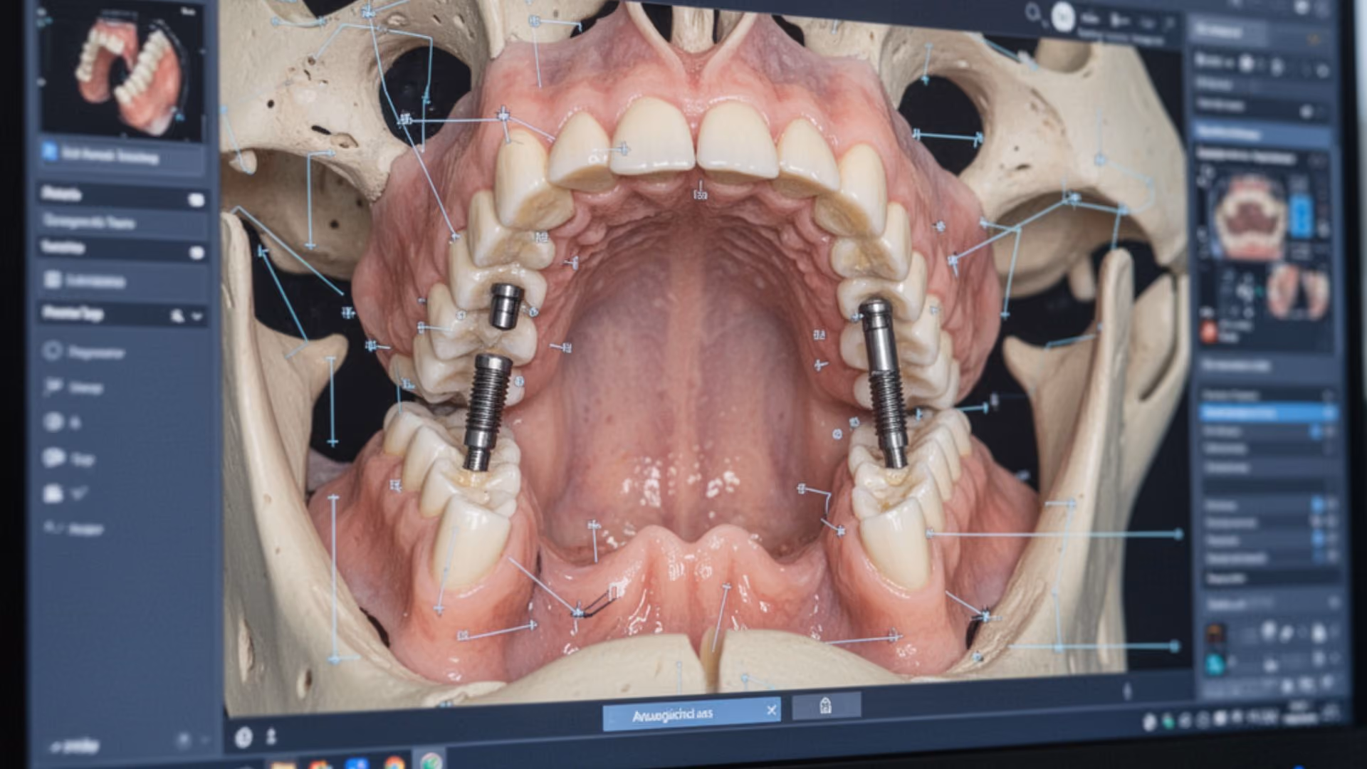
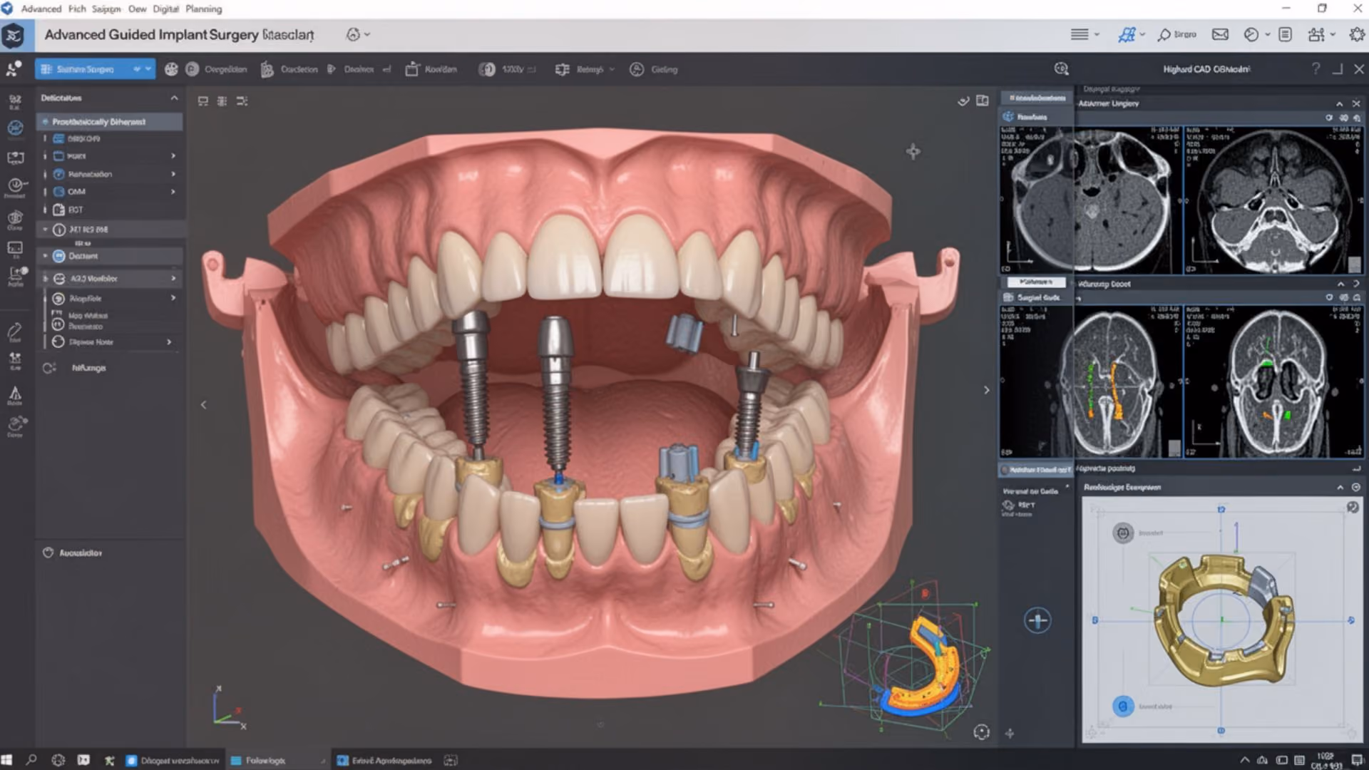
The primary assessment tool is CBCT imaging, which provides a three-dimensional dataset from which bone height, width, and density can be measured at any proposed implant site. This is performed for every implant case at Dazzle before treatment planning is finalised.
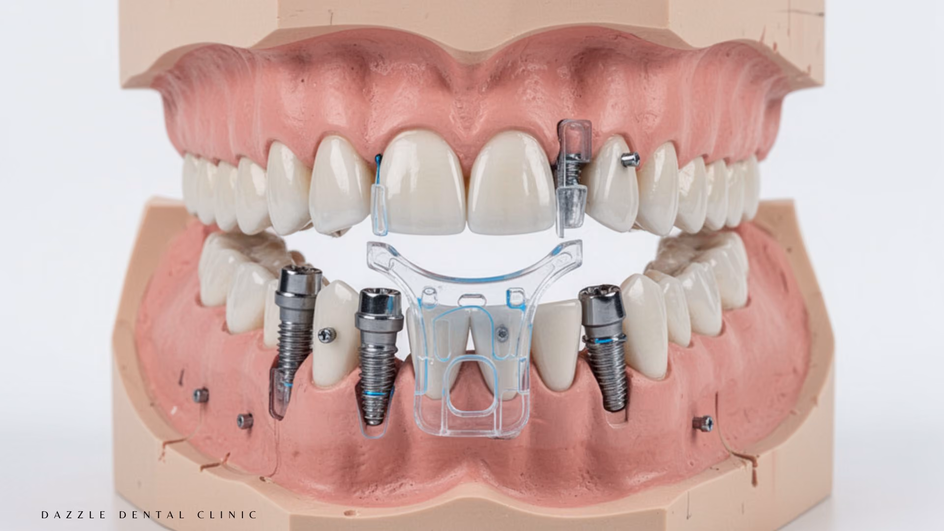
The CBCT data is reviewed by the implantologist alongside the virtual surgical plan — the two are integrated. Implant positions are tested against the actual bone anatomy, and the assessment findings directly modify the plan: a site with insufficient width may receive a narrower implant or be planned for ridge augmentation; a soft-bone posterior maxilla site may prompt a different implant system selection; a site with reduced height above the sinus may be scheduled for a sinus lift procedure before implant placement.
For some cases, a clinical examination component adds relevant information: probing of the gum tissue around remaining teeth reveals the bone level; palpation of the ridge gives a tactile sense of the cortical plate; evaluation of adjacent teeth identifies any periodontal disease that must be resolved before implants are placed.
What Happens When Bone Is Insufficient
Bone deficiency is common and generally manageable. The appropriate response depends on the type and extent of deficiency:
Insufficient width: Guided bone regeneration (GBR) using a membrane and bone graft material can augment the ridge in preparation for implant placement. Healing before implant placement: 4–6 months. Alternatively, narrower-diameter implants may be appropriate if the deficit is minor.
Insufficient height in the posterior maxilla: Sinus lift (sinus floor elevation) creates space for implants beneath the sinus floor. This can be performed simultaneously with implant placement (internal lift, for small deficits) or as a separate staged procedure (lateral window approach, for larger deficits), healing 4–6 months before implant placement.
Low density (soft bone): Protocol modifications at surgery — under-drilling, implant system selection favouring aggressive thread designs — can achieve adequate primary stability in many Type III–IV bone sites without augmentation. For severe cases, alternative anchorage strategies including zygomatic implants bypass the deficient alveolar bone entirely.
Combined deficiency: Full-arch rehabilitation in severely atrophic jaws may involve staged augmentation, immediate extraction and socket grafting, or zygomatic/pterygoid implant protocols. The workup findings determine which applies.
How the Assessment Changes Your Treatment Plan
Many patients arrive at consultation having been told elsewhere that they are “not a candidate” for implants. Occasionally this is accurate. More often, the original assessment was performed from a 2D panoramic without CBCT, without specialist review, or without considering the full range of protocols available for compromised bone.
A comprehensive CBCT-based bone assessment at Dazzle frequently identifies options that were not visible from the original 2D imaging. If conventional implants are viable, we will say so. If augmentation is needed first, we will scope and schedule it. If zygomatic implants are the appropriate solution, we will explain why and what that involves. The clinical findings determine the recommendation — not a default answer.
FAQs
Q1: I was told I don’t have enough bone for implants. Does this mean implants aren’t possible?
Not necessarily. This depends on what type of bone deficiency exists, where it is, and what protocol was considered. Many patients told they cannot have implants are viable candidates for All-on-4 with angled implants (which avoid bone-deficient posterior regions), bone augmentation before implants, or zygomatic implants for severe upper jaw bone loss. A CBCT-based specialist assessment is the only reliable way to determine what’s actually possible.
Q2: How long does bone assessment take at consultation?
The CBCT scan itself takes 10–40 seconds. Review and integration with the treatment plan happens at the consultation appointment. You typically leave the first consultation with a clear understanding of your bone status and the proposed treatment pathway.
Q3: Can bone density improve before surgery?
Not meaningfully through dietary or lifestyle changes in adults. Bone density at a given site is largely fixed by age and systemic health. What can be done is augmenting volume where it is deficient, and selecting surgical protocols appropriate to the existing density. For patients with systemic conditions affecting bone density (osteoporosis, long-term corticosteroid use), the medical management of those conditions before surgery is the relevant optimisation.
Q4: Will my bone continue to change after implants are placed?
Some marginal bone remodelling (typically 1–1.5mm in the first 12–18 months) is normal. After stabilisation, bone levels around well-integrated implants with proper occlusal loading and good hygiene should remain relatively stable for decades. Progressive bone loss beyond the initial remodelling indicates a problem — usually peri-implantitis — that requires assessment and treatment.